17 October 2011 (Monday) - Hypo, Micro...


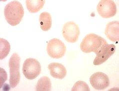
A blood sample on a chap in his mid-forties was received. No clinical details were provided, and there was no previous history.


GONZALES,SPEEDY  DOB  22/10/1967
Sex M Pat No 654321      Source    GP   Received  10:23
Address   SPAIN        Clinician POPL             17/10/2011

Haemoglobin           10.5           g/dl      (13 to 18)
White Blood Cells     7.2            10^9/l    (4 to 11)
Platelets             281            10^9/l    (150 to 400)
Red Blood Cells       5.38           10^12/l   (4.5 to 6)
Haematocrit           0.349          ratio     (0.4 to 0.50)
Mean Cell Volume      64.9           fl        (80 to 100)
Mean Cell Haemoglobin 19.5           pg        (27 to 32)
MCHC                  30.1           g/dl      (32 to 36)
Neutrophils            4.4           10^9/l    (2 to 7.5)
Lymphocytes            2.1           10^9/l    (1.5 to 4)
Monocytes              0.6           10^9/l     (0.2 to 1)
Eosinophils            0.1           10^9/l     (0.02 to 0.5)
Basophils              0.0           10^9/l     (0 to 0.1)
Blood film             ^A blood film has been reviewed


The blood film showed a mild microcytic hypochromic anaemia, target cells, pencil cells and some tear drop cells were seen.


GONZALES,SPEEDY  DOB  22/10/1967
Sex M Pat No 654321      Source    GP   Received  10:23
Address   SPAIN        Clinician POPL             17/10/2011

Sodium                     138  mmol/L   (133 to 146)
Potassium                  5.3  mmol/L   (3.5 to 5.3)
Bicarbonate                24   mmol/L   (22 to 29)
Creatinine                 81   umol/L   (64 to 104)
GFR (estimated)            >90  units=*
Albumin                    42   g/L      (35 to 50)
Total Bilirubin            34 umol/L      (0 to 29)
Alkaline Phosphatase       55 U/L         (30 to 130)
ALT                        13 U/L         (0 to 70)
Calcium                    2.3 mmol/L (   (2.2 to 2.6)
Albumin-corrected calcium  2.3 mmol/L     (2.2 to 2.6)
Phosphate                  1.09 mmol/L    (0.80 to 1.50)

Total Cholesterol          4.1 mmol/L     (2.0 to 5.0)
Fasting sample
HDL Cholesterol            0.9 mmol/L        (0.9 to 1.4)
LDL Cholesterol            2.8  mmol/L 
T.Chol/HDL Ratio           4.6

Iron                       28.6 umol/L (11 to 28)
TSH                        1.5 mIU/L  (0.4 to 5.0)
Free T4                    13  pmol/L (9 to 19)
Serum Vitamin B12          401 ng/L   (189 to 883)
Serum Folate               5.9  ug/L (4.8 to 19.0)


Normally when faced with hypochromia and microcytosis, one thinks of iron deficiency. However the iron levels are fine: if anything a tad high. However serum iron is a transient thing. This should be confirmed by performing a transferring saturation index, or a ferritin assay.
Also the platelet count is not elevated, which often it in with a chronic iron deficiency.

Bearing in mind the elevated red cell count, together with a mildly elevated bilirubin (indicating some increased haemolysis), and the target cells in the blood film, the most likely cause of this blood picture is a thalassaemia trait.
Haemoglobin HPLC should be performed to determine the HbA2 level to distinguish between alpha and beta thalassaemia. Family studies might also be performed.

13 October 2011 (Thursday) - Phaeochromocytoma


A phaeochromocytoma is a rare neuroendocrine tumor of the medulla of the adrenal glands (originating in the chromaffin cells), or extra-adrenal chromaffin tissue that failed to involute after birth and secretes excessive amounts of catecholamines, usually noradrenaline and adrenaline (to a lesser extent).
Extra-adrenal paragangliomas (often described as extra-adrenal pheochromocytomas) are closely related (though less common) tumors that originate in the ganglia of the sympathetic nervous system and are named based upon the primary anatomical site of origin.
The aorticosympathetic group of the adult paraganglia (derived from the embryonic organ of Zuckerkandl) is often a source of paragangliomas.

The signs and symptoms of a pheochromocytoma are those of sympathetic nervous system hyperactivity, including:

  • Skin sensations
  • Flank pain
  • Elevated heart rate
  • Elevated blood pressure, including paroxysmal (sporadic, episodic) high blood pressure, which sometimes can be more difficult to detect; another clue to the presence of pheochromocytoma is orthostatic hypotension (a fall in systolic blood pressure greater than 20 mmHg or a fall in diastolic blood pressure greater than 10 mmHg upon standing)
  • Palpitations
  • Anxiety often resembling that of a panic attack
  • Diaphoresis (excessive sweating)
  • Headaches
  • Pallor
  • Weight loss
  • Localized amyloid deposits found microscopically
  • Elevated blood glucose level (due primarily to catecholamine stimulation of lipolysis (breakdown of stored fat) leading to high levels of free fatty acids and the subsequent inhibition of glucose uptake by muscle cells. Further, stimulation of beta-adrenergic receptors leads to glycogenolysis and gluconeogenesis and thus elevation of blood glucose levels).
 A pheochromocytoma can also cause resistant arterial hypertension. A pheochromocytoma can be acutely fatal if it causes malignant hypertension, or severely high blood pressure. This hypertension is not well controlled with standard blood pressure medications.

Not all patients experience all of the signs and symptoms listed. The most common presentation is headache, excessive sweating, and increased heart rate, with the attack subsiding in less than one hour. Excessive vomitting triggered by an attack will cause compression of the tumour, and therefore release of more hormones, and so exasperating symptoms. It is for this reason that palpitating phaeochromocytomas is not advised.

Tumors may grow very large according to the literature, but nearly all are smaller than 10 cm, most being grape-sized (or smaller)

12 October 2011 (Wednesday) - lingua villosa


Hairy tongue (lingua villosa) is a commonly observed condition of defective desquamation of the filiform papillae that results from a variety of precipitating factors. Usually the ends of the papillae get rubbed away by food but sometimes they grow much longer than normal, making the tongue look furry.

This extra tissue can get stained and become a variety of colours. The condition is most frequently referred to as black hairy tongue (lingua villosa nigra); however, hairy tongue may also appear brown, white, green, pink, or any of a variety of hues depending on the specific etiology and secondary factors (eg, use of coloured mouthwashes, breath mints, tobacco). Bacterial and fungal infections may also complicate the condition.

Whilst dramatic in appearance, use of a toothbrush on the tongue (together with any appropriate antibiotics) is normally enough to effect a cure.

10 October 2011 (Monday) - Diverticulitis


Diverticula is the medical term that is used to describe the small pouches that stick out of the side of the colon. These small pouches are weak spots, much like a bubble forming on a worn inner tube.
Diverticula are very common and associated with ageing. It is estimated that 50% of people have diverticula by the time they are 50 years old, and 70% of people have them by the time they are 80 years old. Although diverticula can occur anywhere in the colon, 80% are found in the lower left side (sigmoid colon) because that is where the colon is the narrowest and the inner pressure the highest.

The majority of people with diverticula will not have any symptoms. However, twenty five per cent of people with diverticula experience symptoms such as abdominal pain and diarrhoea. People who experience symptoms are said to have diverticular disease.
The diverticula can also become infected and inflamed. Inflammation of the diverticula is known as diverticulitis. Diverticulitis causes more severe symptoms than diverticular disease, such as severe pain. It carries a risk of causing serious complications, such as the colon rupturing, which can lead to peritonitis.

 Once suspected, the diagnosis of diverticular disease can be confirmed by a variety of tests. Barium X-rays can be performed to visualize the colon. Diverticula are seen as barium filled pouches protruding from the colon wall.  Direct visualization of the intestine can be done with flexible tubes inserted through the rectum and advanced into the colon. Either short tubes (sigmoidoscopes) or longer tubes (colonoscopes) may be used to assist in the diagnosis and to exclude other diseases that can mimic diverticular disease.

In patients suspected of having diverticular abscess causing persistent pain and fever, ultrasound and CT scan examinations of the abdomen and pelvis can be done to detect collections of pus fluid.
Most patients with diverticulosis have minimal or no symptoms, and do not require any specific treatment. A high fibre diet and fibre supplements are advisable to prevent constipation and perhaps prevent the formation of more diverticula.
Patients with mild symptoms of abdominal pain due to muscular spasm in the area of the diverticula may benefit from anti-spasmodic drugs such as:

·         chlordiazepoxide (Librax),
·         dicyclomine (Bentyl),
·         atropine, scopolamine,
·         phenobarb (Donnatal)
·         hyoscyamine (Levsin).

Some sources also recommend avoidance of nuts, corn, and seeds to prevent complications of diverticulosis.

Whether these dietary restrictions are beneficial is uncertain.

6 October 2011 (Thursday) - Staining for Malaria

A little tip for staining blood smears for malarial parasites: have measured amounts of Giemsa's stain already dispensed into universal containers. Prepare these on a weekly basis. When you need the stain, just top up to the 10ml line with pH7.2 buffer, and it's just that little bit quicker.
And stain the smears using a platen on which the slide hangs upside down and the stain fluid stays in in place by surface tension.
Far less stain deposit, and uses a lot less stain that a traditional Coplin jar full would use.

5 October 2011 (Thursday) - Sickle Cell Disease

Patient presented with abdominal pains early on a Saturday morning

Haemoglobin                6.5   g/dl   (    11 to 15    )
White Blood Cells          11.4  10^9/l (     4 to 11    )
Platelets                  351   10^9/l (   150 to 400   )
Red Blood Cells            2.49  10^12/l(   3.8 to 4.8   )
Haematocrit                0.191 ratio  (  0.36 to 0.46  )
Mean Cell Volume           76.7  fl     (    80 to 100   )
Mean Cell Haemoglobin      26.1  pg     (    27 to 32    )
Mean Cell Haemoglobin Con  34.0  g/dl   (    32 to 36    )
Neutrophils                7.5   10^9/l (     2 to 7.5   )
Lymphocytes                2.4   10^9/l (   1.5 to 4     )
Monocytes                  1.1   10^9/l (   0.2 to 1     )
Eosinophils                0.3   10^9/l (  0.02 to 0.5   )
Basophils                  0.0   10^9/l (     0 to 0.1   )
Reticulocyte count         153.5 10^9/l (    50 to 100   )

Sickle cells ++; some target cells, occasional howell-jolly bodies. neutrophils with toxic granulation.
Referred to consultant haematologist


Biochemistry:

Sodium          139  mmol/L       (   133 to 146   )
Potassium       4.4  mmol/L       (   3.5 to 5.3   )
Creatinine      50   umol/L       (    49 to 90    )
GFR (estimated) >90  units=*                       
Total Protein   62   g/L          (    60 to 80    )
Albumin         34   g/L          (    35 to 50    )
Globulin        28   g/L          (    20 to 35    )
Total Bilirubin 43   umol/L       (     0 to 22    )
Alk Phos        55   U/L          (    30 to 130   )
ALT             18   U/L          (     0 to 50    )


A sickle cell test was found to be positive. Subsequent HPLC showed only a single peak in the HbS position, and no peak in the HbA position.


Final Diagnosis: Patient in Sickle Cell Crisis

1 October 2011 (Saturday) - ALL? - AML !

Patient presented at GP with abdo pain and lack of appetite.


MOUSE,MICKEY                         A+        22/09/2011 u/k
02/02/1951   Male    696969          ITU
                     abdopain not eating drinking..                                              U         LONC
Specimen No   :  AK672862H               Selected Auth Level : E

 HB     13.5   S000 MONO   2.6    S000
 WBC    7.4    S000 EOS    0.4    S000
 PLT    44     S000 BASO   0.1    S000
 RBC    4.52   S000
 HCT    0.415  S000
 MCV    91.8   S000
 MCH    29.9   S000
 MCHC   32.5   S000
 NEUH   0.3    S000
 LYMPH  4.1    S000


The   biochemistry showed:

Sodium           137   mmol/L       (133 to 146) Auth
Potassium         4.1   mmol/L       (3.5 to 5.3) Auth
Creatinine        274   umol/L       ( 64 to 104) Auth
GFR (estimated)   20    units=*                   Auth
Total Protein     70    g/L          ( 60 to 80 ) Auth
Albumin           33    g/L          ( 35 to 50 ) Auth
Total Bilirubin   27    umol/L       (  0 to 29 ) Auth
Alk Phos          324   U/L          ( 30 to 130) Auth
ALT               25    U/L          (  0 to 70 ) Auth
Calcium           2.3   mmol/L       (2.2 to 2.6) Auth
Alb-cor calcium   2.4   mmol/L       (2.2 to 2.6) Auth
Urate             649   umol/L       (200 to 430) Auth
CRP               122   mg/L         (  0 to 10 ) Auth


The blood film showed blast cells, and the case was referred to consultant haematologist
 
CIRCULATING LYMPHOBLASTS . THROMBOCYTOPENIA . NO VISIBLE PLATELET CLUMPS.
NEUTROPENIA. COULD BE NEWLY PRESENTING ALL. MEDICAL TEAM
INFORMED URGENTLY
Subsequent investigations showed:

Coag:

PT             16.4 s            (12 to 16)     Auth
APTT           39.8 s            (22.0 to 35)   Auth
Fibrinogen     5.44 g/L          (1.90 to 4.30) Auth
Thrombin Time  16.9 s            (13 to 20)     Auth
Reptilase time 17.3 s            (14 to 19)     Auth


Serum Total protein  70 g/L          (60 to 80) Auth
Serum Albumin        33 g/L          (35 to 50) Auth
Globulin             37 g/L          (20 to 35) Auth
Electrophoresis           showed the following:                          Auth
    Comments :
     Increased alpha 1 & 2 globulins
     Slight polyclonal increase in gammaglobulins.
     This electrophoretic pattern is consistent with an acute phase
     response.


 Bone marrow was aspirated and scrutinised:

Bone Marrow Investigation:

Report Sequence No.1     22/09/11   09:50     Status : S000

BONE MARROW ASPIRATE AND TREPHINE FROM RPIC EASILY OBTAINED

APARTICULATE HYPERCELLULAR TRAILS
MEGAKARYOCYTES PRESENT WITH SOME HYPOLOBATED FORMS PRESENT.

DIFFERENTIAL BLASTS 80% PRO 1% META 1% ,MYELO 1%
NEUTROPHILS 3% LYMPHOCYTES 4% EOSONOPHILS 2%  ERYTHROID
8%

ERYTHROPOIEISIS AND GRANULOPOIEISIS MARKEDLY REDUCED AN EXCESS OF LYMPHOBLASTS PRESENT ACCOUNTING FOR 80% OF CELLS.

CONSISTENT WITH ACUTE LYMPHOBLASTIC LEUKAEMIA

Reference Laboratory reported on Immunophenotyping and bone marrow:


Results from a gate representing approx. 97% of Total Nucleated Cells (TNC’s):

Immunophenotyping demonstrates a CD34+ cell population representing approx 48% TNC’s with a profile typical of myeloid progenitors (CD13+, CD33-, 1/2CDCD117+, 1/2HLA+, MPO+ and LYSO-).


BONE MARROW ASPIRATE AND TREPHINE EASILY OBTAINED FROM
RPIC

OLIGOPARTICULATE HYPERCELLULAR PARTICLES AND TRAILS VERY FEW MEGAKARYOCYTES SEEN AND HYPOLOBATED

DIFFERENTIAL: BLASTS 80% PROMYELOCYTES 9% MYELOCYTES 5% ERYTHROID 6%

AN EXCESS OF MYELOBLASTS OF TYPE 1 MORPHOLOGY SEEN WITH MARKEDLY REDUCE ERYTHROPOIEISIS AND GRANULOPOIESIS. CONSISTENT WITH A DIAGNOSIS OF ACUTE MYELOID LEUKAEMIA

VERIFIED BY HAEM MDM


The final word on the diagnosis came from the reference laboratory:


Consultant Comments: Type 1 Blasts account for over 90% of TNC’s. Type 2 blasts occasionally seen. No Auer Rods. Blasts are medium-large sized. Consistent with AML (M1) by morphology and Immunophenotyping.


So the final diagnosis was AML (M1). The moral of this story is that in acute leukaemias, the morphology can be misleading. Consultant medical staff can be unsure on the differentiation between myeloblast and lymphoblast. The final diagnosis must be made by flow cytometry which looks for specific cell markers.